Common eye
disease: associated with
acute persistent visual loss
Acute
persistent visual loss
- คือเป็นมา > 24 ชั่วโมง แยกจาก amaurosis fugax (จาก temporary vascular occlusion, neuronal depression [seizure, migraine])
- แบ่งปัญหาเป็น 3 กลุ่ม ได้แก่
- Media (keratitis, corneal edema, hyphema, lens change, vitreous hemorrhage, uveitis)
- Retina (vascular occlusion, retinal detachment, acute maculopathy)
- Neural visual pathway (optic nerve, chiasmal, retrochiasmal visual pathway)
- ซักประวัติ ได้แก่
- Timing: อาจปิดตาข้างดีแล้วเจอโดยบังเอิญ มักไม่ใช่ acute loss
- เป็น 2 ข้าง: สงสัย retrochiasmal visual pathway
- ลักษณะ: เช่น เป็นเฉพาะ peripheral vision สงสัย subtotal retinal detachment, ischemic optic neuropathy, branch retinal vascular occlusion
- ปวดตา ตาแดง: แสดงถึง infection, inflammation
- อาการร่วม เช่น neurological deficit (stroke), N/V (IIOP)
- ประวัติ trauma, ยา (เช่น anticholinergic), contact lens (keratitis), vascular risk (CRAO, CRVO), myopia (retinal detachment), hyperopic (glaucoma), surgery
- Eye exam: VA, EOM, visual fields, pupil + RAPD, IOP, red reflex, slit lamp
- Immediate treatment ใน CRAO, giant cell arteritis, acute-closure glaucoma with IOP > 40 mmHg
- Emergent refer (< 24 ชั่วโมง) ใน infectious keratitis, endophthalmitis, acute retinal necrosis, hyphema, retinal detachment
- Urgent (24-48 ชั่วโมง) ใน non-infectious uveitis, vitreous hemorrhage, acute maculopathy, CRVO, optic neuritis
**ดูเรื่อง glaucoma, endophthalmitis, keratitis, uveitis ในเรื่อง red eye; ดูเรื่อง hyphema, retinal detachment, vitreous hemorrhage ในเรื่อง traumatic eye injury
Central Retinal Artery Occlusion
- มาด้วย acute painless monocular visual loss with retinal ischemia จาก funduscopic exam เป็นหนึ่งในอาการของ ischemic stroke ส่วนใหญ่เกิดจาก carotid artery atherosclerosis
- จะเริ่มเกิด retinal injury ภายใน 100 นาที ยังไม่มีวิธีการรักษาที่ได้ผล แต่เนื่องจาก CRAO มีพยากรณ์โรคที่แย่มาก ในรายที่มาภายใน 24 ชั่วโมง สามารถทดลองรักษาได้ด้วยวิธีดังนี้
- Ocular massage กด 5-15 วินาทีแล้วปล่อยทันที ทำหลายๆครั้ง
- Anterior chamber paracentesis หลังจากให้ยาชาแล้ว ใช้ needle no.30 หันด้านเอียงขึ้น แทงเข้าที่ limbus แล้วดูดน้ำออกจาก AC 0.1-0.2 cc หลังจากนั้นให้ topical ATB
- Acetazolamide 500 mg IV/PO single dose, IV mannitol และ topical medication อื่นๆในการลด IOP
- Vasodilator เช่น pentoxifylline, nitroglycerin, ISDN
- Hyperbaric oxygen therapy ภายใน 2-12 ชั่วโมง
- Carbogen therapy (5% CO2, 95% O2) 10 min q 2 h x 48 h (ช่วยขยาย retinal arterioles)
- Branch retinal artery occlusion (BRAO) มีพยากรณ์โรคดีกว่ามาก ไม่ต้องทำ acute treatment
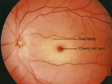 |
| CRAO |
Giant Cell Arteritis/ Temporal arteritis
- เป็นสาเหตุของ CRAO และ ischemic optic neuropathy
- สงสัยในรายที่มีอายุ > 50 ปี + ข้อใดข้อหนึ่ง ได้แก่ new headache (ส่วนใหญ่ที่ temporal, occipital), sudden visual loss, jaw claudication, unexplained fever หรือ anemia, elevated ESR หรือ CRP; โดยเฉพาะในคนที่มีประวัติเป็น polymyalgia rheumatica
- methylprednisolone 1000 mg IV OD x 3 วัน ใน acute visual loss รวมถึง amaurosis fugax, diplopia หรือ visual dysfunction อื่นๆ; + aspirin 80-100 mg/d + PPI หรือ misoprostol
 |
| temporal arteritis |
Acute retinal
necrosis
- ส่วนใหญ่เกิดจาก VZV มาด้วย acute iridocyclitis, vitritis, necrotizing retinitis, occlusive retinal vasculitis, retinal detachment มีอาการตามัว ปวดตา
- ให้ IV acyclovir
Retinal Detachment/ retinal tear
- แบ่งตามสาเหตุเป็น rhegmatogenous (retina break), nonrhegmatogenous (leak, exudative retinal detachment), vitreous traction (traction retinal detachment)
- ถ้าเกิดตามหลัง posterior vitreous detachment มักจะมาด้วยมี floater เหมือนใยแมงมุมลอยไปมา อาจมีแสงวูบวาบ (photopsia) จุดดำลอยไปมา (vitreous hemorrhage) และมี peripheral visual field defect
- Refer พบ retina specialist ภายใน 24 ชั่วโมง ถ้ามี macula-on retinal detachment
Optic
neuropathy
- ในคนอายุน้อย (18-40) มักเกิดจาก optic neuritis (สัมพันธ์กับ multiple sclerosis) ส่วนในคนอายุมาก (> 50) มักเกิดจาก ischemic optic neuropathy
- มาด้วย monocular visual loss (painful ใน optic neuritis; painless ใน optic ischemic neuropathy), เป็นแบบ central vision loss (scotoma), RAPD present, dyschromatopsia (มองเห็นสีเปลี่ยนไป), optic disc swelling
- ในรายที่สงสัย optic neuritis ให้ตรวจ Brain and orbit MRI with Gd รักษาโดยให้ IV methylprednisolone
- ให้คิดถึงสาเหตุจาก giant cell arteritis เสมอ ซึ่งมักพบในคนอายุ > 70 ปี (ดูด้านบน)
Retinal Vein Occlusion
- แบ่งเป็น BRVO (branch), CRVO (central), HRVO (hemi)
- มักจะไม่มีอาการ อาจมีตามัวได้ อาจมี relative RAPD ได้ ตรวจพบ focal หรือ wedge-shaped retinal hemorrhage มี apex ตำแหน่งที่ AV crossing
- Urgent consultation
 |
| CRVO |
ไม่มีความคิดเห็น:
แสดงความคิดเห็น